44 cell diagram with labels
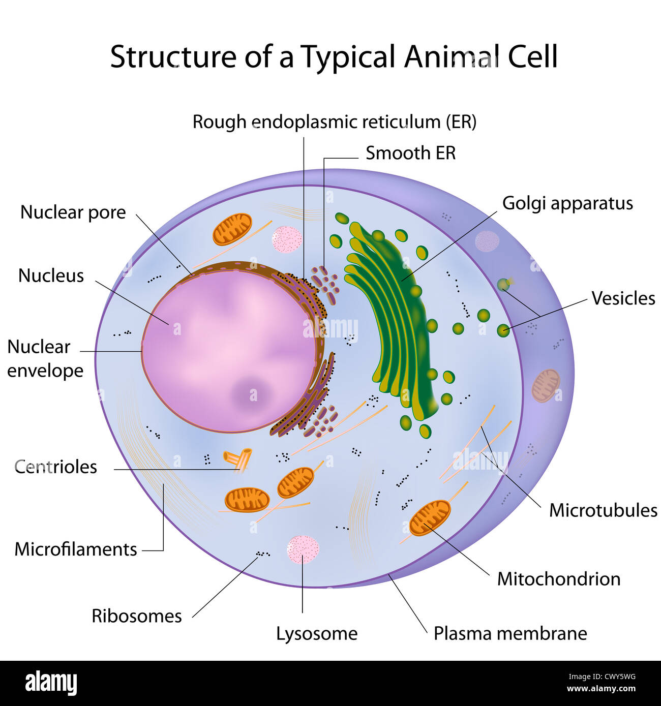
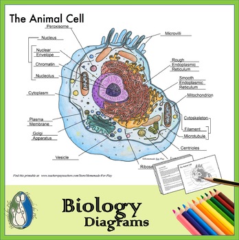
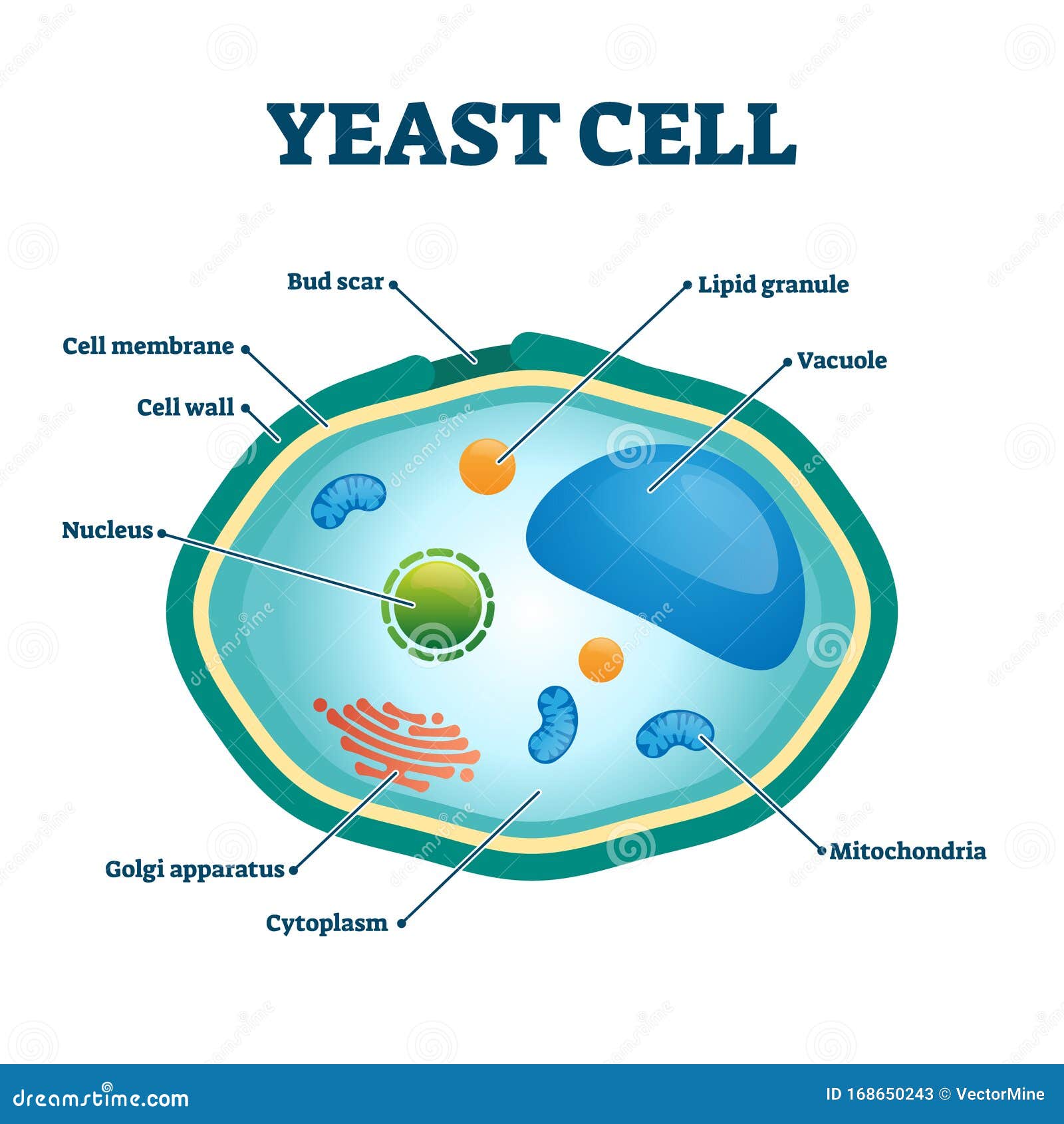
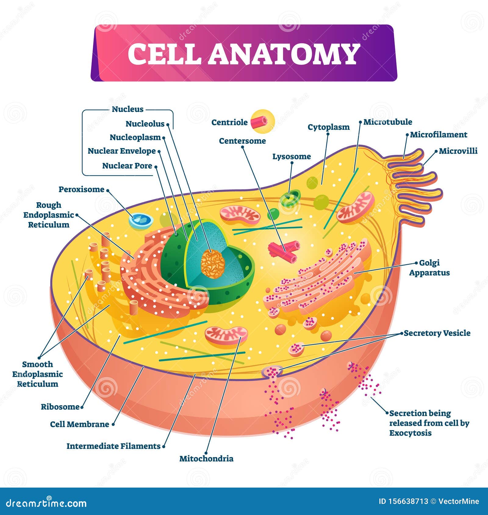
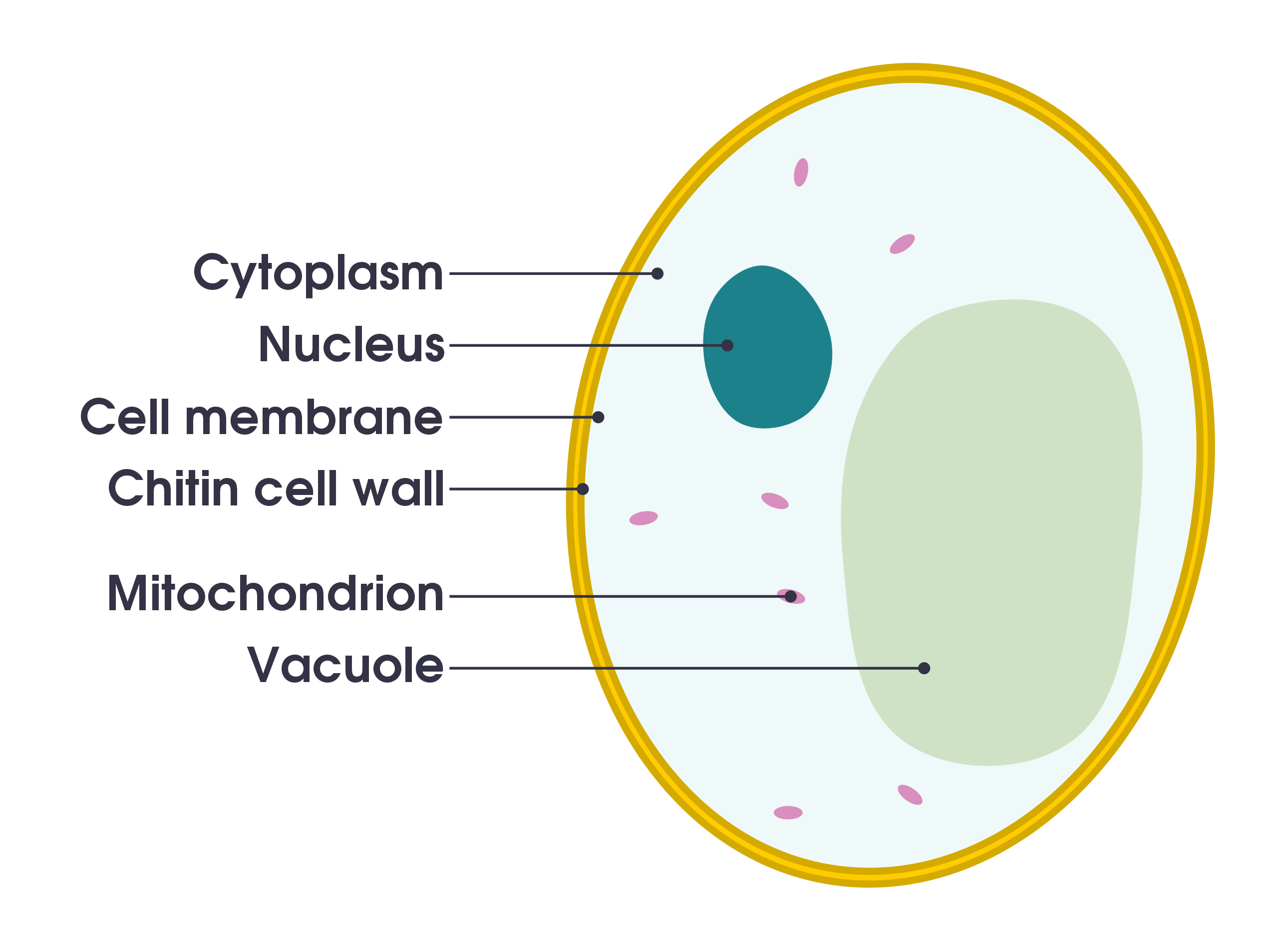
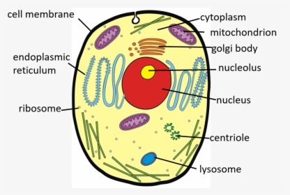
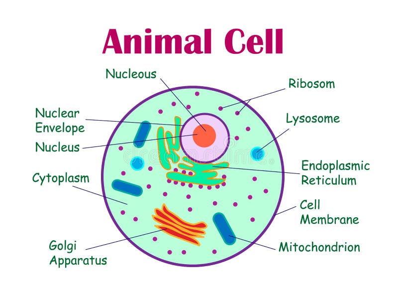
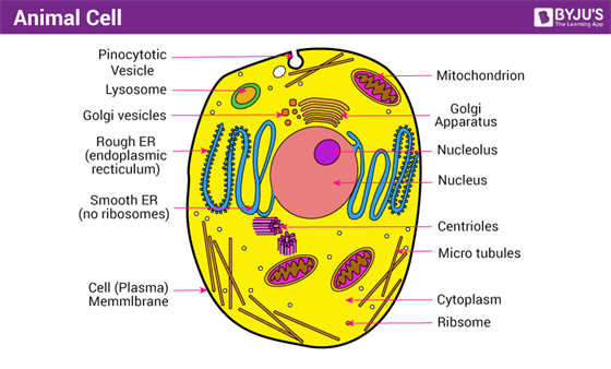
Cell: Structure and Functions (With Diagram) - Biology Discussion Eukaryotic Cells: 1. Eukaryotes are sophisticated cells with a well defined nucleus and cell organelles. 2. The cells are comparatively larger in size (10-100 μm). 3. Unicellular to multicellular in nature and evolved ~1 billion years ago. 4. The cell membrane is semipermeable and flexible. 5. These cells reproduce both asexually and sexually. A Well-labelled Diagram Of Animal Cell With Explanation - BYJUS The animal cell diagram is widely asked in Class 10 and 12 examinations and is beneficial to understand the structure and functions of an animal. A brief explanation of the different parts of an animal cell along with a well-labelled diagram is mentioned below for reference. Also Read Different between Plant Cell and Animal Cell
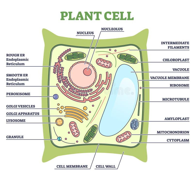
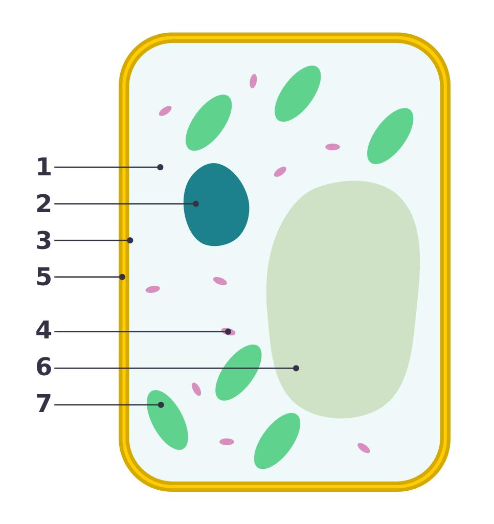
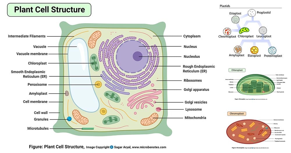
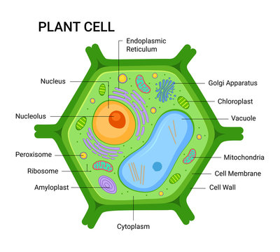
Plant and Animal Cell: Labeled Diagram, Structure, Function - Embibe Plant Cell: Plant cells are eukaryotic cells with a true nucleus along with specialized structures called organelles that carry out certain specific functions. Animal Cell: An animal cell is a type of eukaryotic cell that lacks a cell wall and has a true, membrane-bound nucleus along with other cellular organelles. Diagram of Plant and Animal Cell
Cell diagram with labels
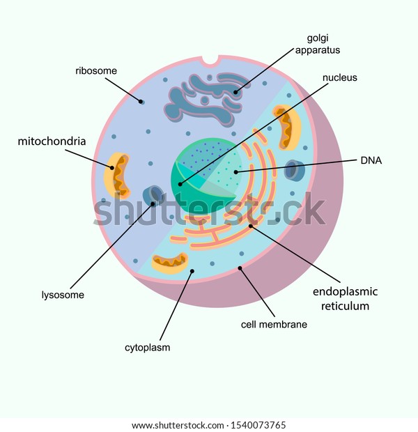
Human Cell Diagram, Parts, Pictures, Structure and Functions Diagram of the human cell illustrating the different parts of the cell. Cell Membrane. The cell membrane is the outer coating of the cell and contains the cytoplasm, substances within it and the organelle. It is a double-layered membrane composed of proteins and lipids. The lipid molecules on the outer and inner part (lipid bilayer) allow it to ... Cell Organelles- Definition, Structure, Functions, Diagram - Microbe Notes Animal Cell- Definition, Structure, Parts, Functions, Labeled Diagram Prokaryotes vs Eukaryotes- Definition, 47 Differences, Structure, Examples Amazing 27 Things Under The Microscope With Diagrams Structure of Cell: Definition, Types, Diagram, Functions - Embibe Various kinds of cells show special differences, yet they all have some basic structural plan consisting of three essential parts: (i) cell membrane ( plasma membrane ), (ii) cytoplasm and (iii) nucleus. Apart from these three components, cells have some living parts that are called cell organelles.
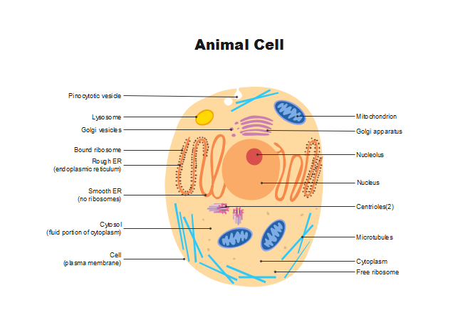
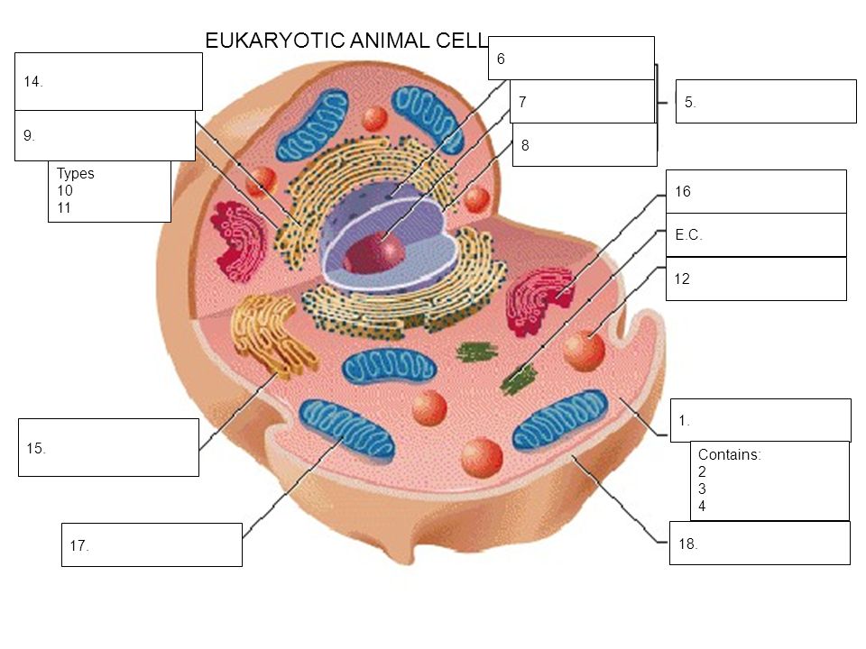
Cell diagram with labels. Animal Cells: Labelled Diagram, Definitions, and Structure - Research Tweet The endoplasmic reticulum (s) are organelles that create a network of membranes that transport substances around the cell. They have phospholipid bilayers. There are two types of ER: the rough ER, and the smooth ER. The rough endoplasmic reticulum is rough because it has ribosomes (which is explained below) attached to it. Learn the parts of a cell with diagrams and cell quizzes For this exercise we'll start with an image of a cell diagram ready labeled. Study this and make sure that you're clear about which structure is found where. Cell diagram unlabeled It's time to label the cell yourself! As you fill in the cell structure worksheet, remember the functions of each part of the cell that you learned in the video. Labeled Plant Cell With Diagrams | Science Trends The parts of a plant cell include the cell wall, the cell membrane, the cytoskeleton or cytoplasm, the nucleus, the Golgi body, the mitochondria, the peroxisome's, the vacuoles, ribosomes, and the endoplasmic reticulum. Parts Of A Plant Cell The Cell Wall Let's start from the outside and work our way inwards. Cell Diagram | Free Cell Diagram Templates - Edrawsoft Cell Diagram Template A free customizable cells diagram template is provided to download and print. Quickly get a head-start when creating your own cell diagram. Here is a simple cell diagram example created by Science Diagram Maker Download Template: Get EdrawMax Now! Free Download Popular Latest Flowchart Process Flowchart Workflow BPMN
Label the cell diagram - Teaching resources - Wordwall by Mercedkeishla. Label Plant and Animal Cell Labelled diagram. by Arucker. G9 Biology. English/Spanish Label the Diagram! (auto, graph, phon, photo, tele, logy) Labelled diagram. by Alissaccasey. 8.1 Label the sentence Labelled diagram. by Christianjolene. 5.7 Label the sentence Labelled diagram. 2,141 Red blood cell diagram Images, Stock Photos & Vectors | Shutterstock 2,141 red blood cell diagram stock photos, vectors, and illustrations are available royalty-free. See red blood cell diagram stock video clips Image type Orientation Color People Artists More Sort by Popular Biology Science Geography and Landscapes Healthcare and Medical red blood cell blood vessel blood oxygen hemoglobins medicine Next of 22 Plant Cell Diagram | Science Trends A plant cell diagram, like the one above, shows each part of the plant cell including the chloroplast, cell wall, plasma membrane, nucleus, mitochondria, ribosomes, etc. A plant cell diagram is a great way to learn the different components of the cell for your upcoming exam. Plants are able to do something animals can't: photosynthesize. Label that Diagram - Cells - Apps on Google Play There are 5 cells presented: Animal Cell, Plant Cell, Amoeba, Paramecium, and Euglena. The player can study the labeled diagrams or play the game of labeling the diagrams. When the game is played, the labels appear in a random order one at a time and the player must tap on the correct dot on the diagram.
Label Cells Teaching Resources | Teachers Pay Teachers Animal & Plant Cell: Label the Diagram and Differences table:This is a great supplement for students to review/assess and strengthen their knowledge the unit of ANIMAL AND PLANT CELL UNIT. Answer key included.It includes total TWO worksheets. This worksheets can demonstrates relationships between facts and concepts.It can be used as a small ... Label Cell Parts | Plant & Animal Cell Activity | StoryboardThat Click "Start Assignment". Find diagrams of a plant and an animal cell in the Science tab. Using arrows and Textables, label each part of the cell and describe its function. Color the text boxes to group them into organelles found in only animal cells, organelles found in only plant cells, and organelles found in both cell types. cell diagrams to label Plant Cell Diagram With Labels And Functions - Diagram Media. 17 Pics about Plant Cell Diagram With Labels And Functions - Diagram Media : Animal Cell No Label - Plant Cell Diagram Without Labels Plant Cell, draw the diagrams of different types of cells and label them - Brainly.in and also Plant Cell Diagram And Label Simple - Cell Diagram. Labeling a Cell Diagram | Quizlet Cell Wall This gives shape and support to the plant cell. It surrounds the cell and protects the other parts of the cell. Chloroplasts This is where the plant cell's chlorophyll is stored. This is what the plant uses to make its own food (photosynthesis). This is also what makes plant cells have a green-like color. Plant cells Are circular in shape
03 Label the Cell Diagram | Quizlet Start studying 03 Label the Cell. Learn vocabulary, terms, and more with flashcards, games, and other study tools.
Cell Diagrams with Labelling Activity | Learnful I've created two interactive diagrams for an upcoming open textbook for high-school level biology. The cell structure illustrations for these diagrams were generated in BioRender. Both diagrams feature a drag-and-drop labelling activity created with H5P here on Learnful. These h5p resources are made available openly with the CC BY license.
A Labeled Diagram of the Animal Cell and its Organelles A Labeled Diagram of the Animal Cell and its Organelles There are two types of cells - Prokaryotic and Eucaryotic. Eukaryotic cells are larger, more complex, and have evolved more recently than prokaryotes. Where, prokaryotes are just bacteria and archaea, eukaryotes are literally everything else.
PDF Human Cell Diagram, Parts, Pictures, Structure and Functions Diagram of the human cell illustrating the different parts of the cell. Cell Membrane The cell membraneis the outer coating of the cell and contains the cytoplasm, substances within it and the organelle. It is a double-layered membrane composed of proteins and lipids.
Plant cell diagrams: label parts Sixth grade Science Worksheets Free questions for "Plant Cell Diagrams: Label Parts" to improve science understanding and many other skills. Science worksheets perfect for 6th-grade students. Categories Science, Sixth grade Post navigation. Body systems: perception and motion Sixth grade Science Worksheets.
cell diagram to label nucleus ribosomes diagram structure labeled cell biology eukaryotic cells which khan 2.3.1 Draw And Label A Diagram Of The Ultrastructure Of A Liver Cell As liver cell drawing diagram draw label ultrastructure paintingvalley animal example labelling diagrams ze kig source Quia - Chapter 3: Bacteria & Protists quia.com
Cell Organelles Label Teaching Resources | Teachers Pay Teachers This worksheet is designed to help students bring together all of the interconnected organelles in the cell. Students read statements and label the statements with a number from the diagram. In the diagram, students will see how substances are transported between the nucleus, endoplasmic reticulum, golgi, lysosomes, vesicles, and food vacuoles.
Free Cell Diagram Software with Free Templates - EdrawMax - Edrawsoft Before making a cell diagram on EdrawMax, first gather all the necessary supporting facts to draw the diagram. Draw all the cell components roughly into the shape of a cell. The cell wall, cell membrane, cytoplasm, nucleus, and cell organelles are components. Step 2: Template selection. Step 3: Customize the Diagram.
Converting Diagrams - The Biology Corner Open Google Draw and import the diagram. Then use "insert" to create text boxes where students can fill in the labels. Don't forget when assigning this to students on Google classroom to make a copy for each student. You can leave documents in an uneditable form and students can use an addon like "Kami" to annotate the document.
bone cell diagram with labels bone cell diagram with labels Skeletal system human anatomy chart skeleton labeled diagram structure anatomical body 1000 sketch bones bone parts labels organs complete joint. Anatomy and physiology questions.
Animal Cell Diagram with Label and Explanation: Cell ... - Collegedunia Animal cell is a typical Eukaryotic cell enclosed by a plasma membrane containing nucleus and organelles which lack cell walls, unlike all other Eukaryotic cells. The typical cell ranges in size between 1-100 micrometers. The lack of cell walls enabled the animal cells to develop a greater diversity of cell types.
Structure of Cell: Definition, Types, Diagram, Functions - Embibe Various kinds of cells show special differences, yet they all have some basic structural plan consisting of three essential parts: (i) cell membrane ( plasma membrane ), (ii) cytoplasm and (iii) nucleus. Apart from these three components, cells have some living parts that are called cell organelles.
Cell Organelles- Definition, Structure, Functions, Diagram - Microbe Notes Animal Cell- Definition, Structure, Parts, Functions, Labeled Diagram Prokaryotes vs Eukaryotes- Definition, 47 Differences, Structure, Examples Amazing 27 Things Under The Microscope With Diagrams
Human Cell Diagram, Parts, Pictures, Structure and Functions Diagram of the human cell illustrating the different parts of the cell. Cell Membrane. The cell membrane is the outer coating of the cell and contains the cytoplasm, substances within it and the organelle. It is a double-layered membrane composed of proteins and lipids. The lipid molecules on the outer and inner part (lipid bilayer) allow it to ...

















![1.1: Structure of the generalized cell [27]. | Download ...](https://www.researchgate.net/profile/Aggrey-Wakhule/publication/325897026/figure/fig1/AS:639921136091136@1529580496082/1-Structure-of-the-generalized-cell-27.png)


Post a Comment for "44 cell diagram with labels"